Call us
+355697772828
+355697772828
The key to a successful treatment starts with an accurate diagnosis. Without a complete diagnosis, the patient faces not only an incomplete treatment but also an incorrect one. We possess the most advanced European equipment with the lowest radiation exposure.
We analyze all types of X-rays in detail to make an accurate diagnosis, without errors:

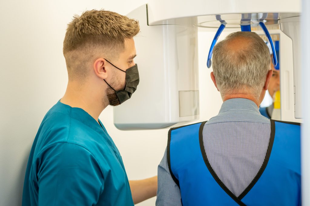
At A S C, you will find a computed tomography scanner (3D Scanner). This is a progressive and advanced diagnostic method that ensures precision in measurements down to a fraction of a millimeter. With the help of computed tomography in 3D mode, the diagnosis of cavities and processes in tissues, which are difficult to detect with other methods, is made possible.
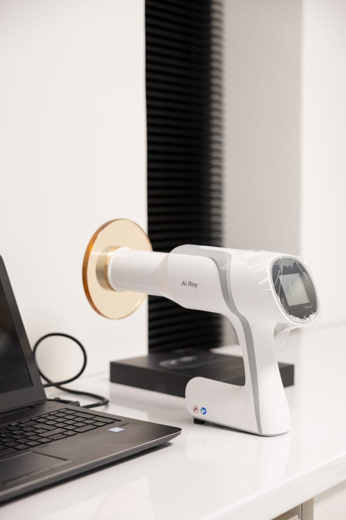
The dental visiograph (also known as a radiovisiograph) is a modern diagnostic device that allows patient examination during treatment, directly from the dental chair. The visiographic examination method assists the dentist in detecting even the smallest dental pathologies with greater precision.
The visiograph converts X-ray radiation into a digital image and transmits it to a computer monitor located at the treatment site. In visiograph diagnostics, the radiation dose for both the patient and the dentist is ten times lower compared to a conventional X-ray device.

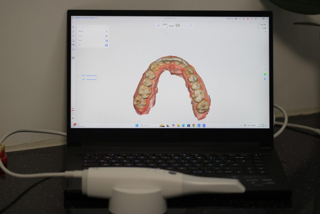
The intraoral scanner in dentistry is a modern device used to create three-dimensional models of a patient’s teeth and jaws. It is useful for diagnosis, treatment planning, and monitoring outcomes, providing accurate information about the shape and size of teeth as well as their position relative to one another. The digital models fully replicate jaw movements, enabling dentists to develop the most effective treatment plans.
Scanners are commonly used for planning and performing dental implant procedures and for producing surgical guides. In prosthodontics, they are utilized to fabricate veneers, crowns, bridges, and various other prostheses.

Copyright © 2024 Art Smile Clinic, All rights reserved. Powered by SmartWeb